Mechanical Ventilation:
The Roman physician Galen may
have been the first to describe mechanical ventilation: "If you take a
dead animal and blow air through its larynx [through a reed], you will fill its
bronchi and watch its lungs attain the greatest distention."
A mechanical ventilator is a machine that
generates a controlled flow of gas into a patient’s airways. Oxygen and air are
received from cylinders or wall outlets (internal Gases Network), the gas is
pressure reduced and blended according to the prescribed inspired oxygen
tension (FiO2), accumulated in a receptacle within the machine, and delivered
to the patient using one of many available modes of ventilation.
But before going in depth of Ventilation modes
and Ventilators we should know more about structure of our Respiratory System.

Respiratory System:
The respiratory system provides the means for
gas exchange required in living cells. Examples: Carbon Dioxide and Oxygen.
When you inhale you are bringing oxygen to the lungs. When you exhale you are
getting rid of carbon dioxide. When you hold your breath, does your body start
saying breath I need oxygen or does it say help the carbon dioxide levels that
are out of whack? If you said carbon dioxide levels you are correct. Let's take
a look now at what the respiratory system consists.
There are 2 tracts of the respiratory system:
- Upper respiratory system.
- Lower respiratory system
It also
can be divided into a conducting portion and respiratory function.
Conducting portion: Nose,
Nasal cavity, pharynx, larynx, trachea and the smaller progressively airways
(primary bronchi to terminal bronchioles).
Respiratory portion: is composed of small airways called respiratory
branchioles, alveolar ducts and alveoli (air sacs).
Some of Respiratory System main Functions:

.JPG)
Pressure Controlled (pressure limited, pressure targeted) and Volume Variable.
Dual Controlled (volume targeted (guaranteed) pressure limited).
 |
| Respiratory System |
 |
| Respiratory System Anatomy |
Some of Respiratory System main Functions:
Pulmonary Ventilation:
Inhalation: bringing gas into the lungs.
Exhalation: letting gas flow out of the
lungs.
Gas exchange: Oxygen is drawn in by inhalation and is
transported to the body cells from the lungs by blood circulation. The body
uses the oxygen to generate carbon dioxide as a waste product which is then
transported to the lungs and is then exhaled.
Gas Conditioning: Gases entering the body are
"modified" before reaching the gas exchange surfaces. These gases are
warmed to body temperature, filtered of any harmful particles and humidified by
contact of the respiratory epithelium and the sticky mucus covering in the winding
pathways in the nasal cavity and the paranasal sinuses.
Some Ventilation Concepts:
Negative-pressure ventilation: where air is essentially sucked into the lungs.
Positive pressure ventilation: where air (or
another gas mix) is pushed into the trachea.
When physicians use a ventilator?
Medical Ventilator is a machine designed to mechanically
assist in moving breathable air into and out of the lungs, to provide the
mechanism of breathing for a patient who is physically unable to breathe, or
breathing insufficiently.
Ventilator mainly in its simplest form, a
modern positive pressure ventilator consists of:
- A compressible air reservoir or turbine,
- Air and oxygen supplies,
- A set of valves and tubes,
- A disposable or reusable "patient circuit",
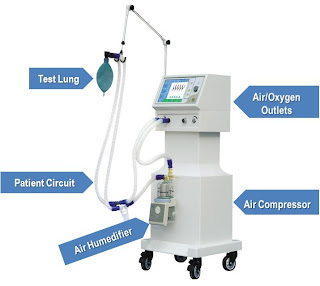

The air reservoir is pneumatically compressed
several times a minute to deliver room-air, or in most cases, an air/oxygen
mixture to the patient. If a turbine is used, the turbine pushes air through
the ventilator, with a flow valve adjusting pressure to meet patient-specific
parameters. When overpressure is released, the patient will exhale passively
due to the lungs' elasticity, the exhaled air being released usually
through a one-way valve within the patient circuit called the patient manifold.
The oxygen content of the inspired gas can be set from 21 percent (ambient air)
to 100 percent (pure oxygen). Pressure and flow characteristics can be set
mechanically or electronically.
Ventilators may also be equipped with
monitoring and alarm systems for patient-related parameters (e.g. pressure,
volume, and flow) and ventilator function (e.g. air leakage, power failure, and
mechanical failure), backup batteries, oxygen tanks, and remote control. The
pneumatic system is nowadays often replaced by a computer-controlled turbo
pump.
Modern ventilators are electronically
controlled by a small embedded system to allow exact adaptation of
pressure and flow characteristics to an individual patient's needs. Fine-tuned
ventilator settings also serve to make ventilation more tolerable and
comfortable for the patient. In Germany, Canada, and the United
States, respiratory therapists are responsible for tuning these
settings while biomedical technologists are responsible for the maintenance.
The patient circuit usually consists of a set
of three durable, yet lightweight plastic tubes, separated by function (e.g.
inhaled air, patient pressure, exhaled air). Determined by the type of
ventilation needed, the patient-end of the circuit may be either noninvasive or
invasive.
Noninvasive methods, which are adequate for
patients who require a ventilator only while sleeping and resting, mainly
employ a nasal mask. Invasive methods require intubation, which for
long-term ventilator dependence will normally be
a tracheotomy cannula, as this is much more comfortable and practical
for long-term care than is larynx or nasal intubation.
Setup of Ventilator in hospital ICU room:


Setup of Ventilator in hospital ICU room:
The illustration shows a standard setup for a
ventilator in a hospital room. The ventilator pushes warm, moist air (or air
with increased oxygen) to the patient. Exhaled air flows away from the patient.
Some expressions:
Minute Volume: is the volume of air (oxygen) inhaled (inhaled
minute volume) or exhaled (exhaled minute volume) from a person’s
lungs in one minute (5-40 L/min).
Tidal Volume (Vt): the volume of air (oxygen) what is given to
patient in one breathe (0.1-2 L).
Flow:
volume/time (5-35 L/min).
BPM: Breathe Per
Minute (2-60)
Ti: Inspiration
time.
Te: Expiration
time.
I:E: Inspiration
to Expiration ratio.
Respiratory Cycle:

Relation between Tidal Volume, Inspiration time
and Flow:

.JPG)
How to work on ventilator? The classification of ventilators refers to the
following elements:
1- Control: How the
ventilator knows how much flow to deliver
Volume Controlled (volume limited, volume targeted) and Pressure
Variable.Pressure Controlled (pressure limited, pressure targeted) and Volume Variable.
Dual Controlled (volume targeted (guaranteed) pressure limited).
2- Cycling: how the
ventilator switches from inspiration to expiration: the flow has been delivered
to the volume or pressure target - how long does it stay there?
Time cycled such in pressure controlled ventilation.
Flow cycled such as in pressure support.
Volume cycled the ventilator cycles to expiration once a set tidal volume has
been delivered: this occurs in volume controlled ventilation. If an inspiratory
pause is added, then the breath is both volume and time cycled.
3- Triggering: what causes the ventilator to cycle to inspiration. Ventilators
may be time triggered, pressure triggered or flow triggered.
Time: the
ventilator cycles at a set frequency as determined by the controlled rate.
Pressure: the ventilator senses the patient's inspiratory effort by way of a
decrease in the baseline pressure.
Flow: modern ventilators
deliver a constant flow around the circuit throughout the respiratory cycle
(flow-by). A deflection in this flow by patient inspiration is monitored by the
ventilator and it delivers a breath. This mechanism requires less work by the
patient than pressure triggering.
4- Breaths are either: what causes the ventilator to cycle from
inspiration
Mandatory (controlled) - which is determined by the respiratory rate.
Assisted (as
in assist control, synchronized intermittent mandatory ventilation, pressure
support)
Spontaneous (no additional assistance in inspiration, as in CPAP)
5- Flow pattern:
Sinusoidal: this is the flow pattern
seen in spontaneous breathing and CPAP
Decelerating: the flow pattern seen in pressure targeted ventilation:
inspiration slows down as alveolar pressure increases (there is a high initial
flow). Most intensives and respiratory therapists use this pattern in volume
targeted ventilation also, as it results in a lower peak airway pressure than
constant and accelerating flow, and better distribution characteristics.
Constant: flow continues at a constant rate until the set tidal volume is
delivered.
Accelerating: flow increases progressively as the
breath is delivered. This should not be used in clinical practice.
6- Mode or Breath Pattern:
Continuous Mandatory Ventilation (CMV): In which ventilator provides a mechanical
breath on a preset timing, without allowances for spontaneous breathing from patient
side. This is usually only used in an unconscious patient. It may also be used
in infants who often quickly adapt their breathing pattern to the ventilator
timing.
Assist-Control: That minimizes
patient effort by providing full mechanical support with every breath. This is
often the initial mode chosen for adults because it provides the greatest
degree of support.
Intermittent Mandatory Ventilation (IMV): It mixes controlled breaths and spontaneous
breaths, as the ventilator provides a preset mechanical breath (volume limited)
every specified number of seconds (determined by dividing the respiratory rate
into 60 seconds, thus a respiratory rate of 12 results in a 5 second cycle
time). Within that cycle time the ventilator waits for the patient to initiate
a breath using either a pressure or flow sensor. When the ventilator senses the
first patient breathing attempt within the cycle, it delivers the preset
ventilator breath. If the patient fails to initiate a breath, the ventilator
delivers a mechanical breath at the end of the breath cycle. Additional
spontaneous breaths after the first one within the breath cycle do not trigger
another SIMV breath. However.
SIMV is frequently employed as a method of
decreasing ventilatory support (weaning) by turning down the rate, which
requires the patient to take additional breaths beyond the SIMV triggered
breath.
Pressure Support: Where the patient has control over all aspects
of his breath except the pressure limit.
Continuous Positive Airway Pressure (CPAP): A continuous level of elevated pressure is
provided through the patient circuit to maintain adequate oxygenation, decrease
the work of breathing, and decrease the work of the heart (such as in left-sided
heart failure CHF). Note that no cycling of ventilator pressures occurs and the
patient must initiate all breaths. In addition, no additional pressure above
the CPAP pressure is provided during those breaths
Synchronized Intermittent Mandatory Ventilation
(SIMV): In which ventilator provides a preset
pressure limited mechanical breath every specified number of seconds SIMV is
frequently employed as a method of decreasing ventilatory support (weaning) by
turning down the rate, which requires the patient to take additional breaths
beyond the SIMV triggered breath.
Positive End Expiratory Pressure (PEEP): may or may not be employed to prevent atelectasis in adult
patients. It is almost always used for pediatric and neonatal patients due to
their increased tendency for atelectasis.
Dräger Company:

As an international leader in medical and
safety technology, Dräger develops innovative equipment and solutions people
the world over trust when livesare on the line and “Technology for Life” is
their guiding principle and mission.
And we invited "Dräger International Co." agent company in Egypt "Life care Technology Co." and their representative Eng. Shawkat Ahmed (Service Supervisor of Co.) who welcomed the invitation and spent about three hours illustrating the Medical Ventilation Machines to SBME's and bring one of their most advanced models of their Ventilators Evita XL (which is shown among their products below).
And we invited "Dräger International Co." agent company in Egypt "Life care Technology Co." and their representative Eng. Shawkat Ahmed (Service Supervisor of Co.) who welcomed the invitation and spent about three hours illustrating the Medical Ventilation Machines to SBME's and bring one of their most advanced models of their Ventilators Evita XL (which is shown among their products below).
Dräger’s range of Ventilators Products:
 |
| Drager - Evita 2 dura |
 |
| Drager - Savina |
 |
| Drager - Evita XL |
 |
| Drager - Oxylog 1000 |
 |
| Drager - Oxylog 2000 |
“We enable professional caregivers
turn the ICU into a healing environment”
–Stefan Draeger.
References:
Operation Manual of Evita XL Ventilator
Medical Ventilation through Wikipedia
Medical Ventilators through Wikipedia
Weaning patient from mechanical ventilator
What To Expect While on a Ventilator?
Modes of Mechanical Ventilation


